Anatomy of the Hoof
There’s been a lot of interest in topics surrounding hoof care lately. It’s a vitally important part of the anatomy, for as the saying goes “No Hoof, No Horse.” Out of an unfortunate necessity, I had to learn more than I ever wanted to know on the subject.
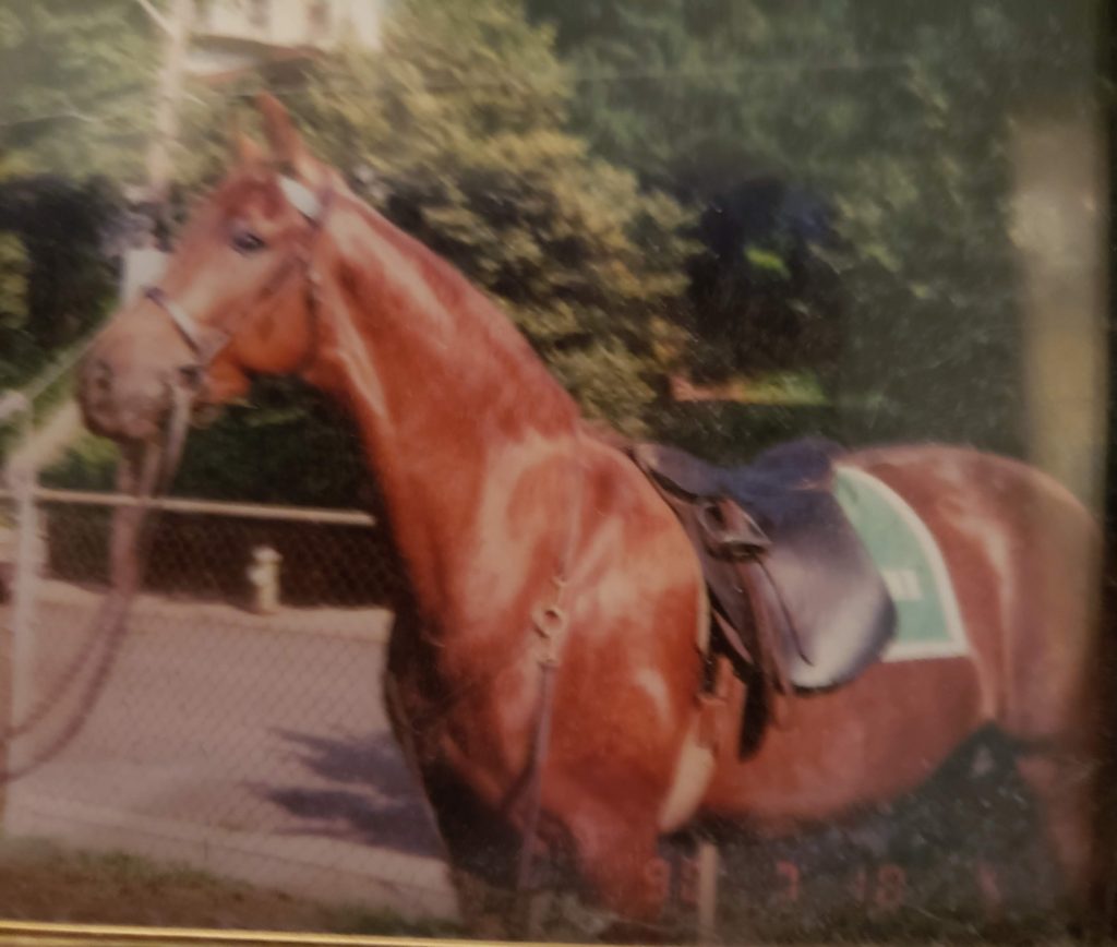
My first horse, “Wimsey” an American Saddlebred ,was involved in a terrible accident on the way to a show. After numerous surgeries, he returned home, and as an answer to my prayers we again enjoyed many wonderful rides together. As time went on, the after effects of the injuries he sustained caused him to founder. That started us on a dark downward spiral that made it necessary for me to learn more about the hoof than I ever thought I would want or need to know.
Many of you may have in-depth knowledge of the hoof. But if not, I’m continuing a series on the subject …a “101”…introductory for some, and an overview refresher for those like myself.
Outer Structures
Hoof Wall
The first part of the hoof is called the hoof wall. This is the hard outer covering that houses and protects the structures within, supports the weight of the horse and acts as a shock absorber as the horse moves. The hoof wall does not have nerves or blood vessels. It’s made up of keratin, a fibrous structural protein, the same thing that makes up hair, nails, and the outer layer of human skin. It grows continuously, and needs to be trimmed if not naturally worn off. A healthy hoof wall grows about 3/8 of an inch per month.
Coronary Band
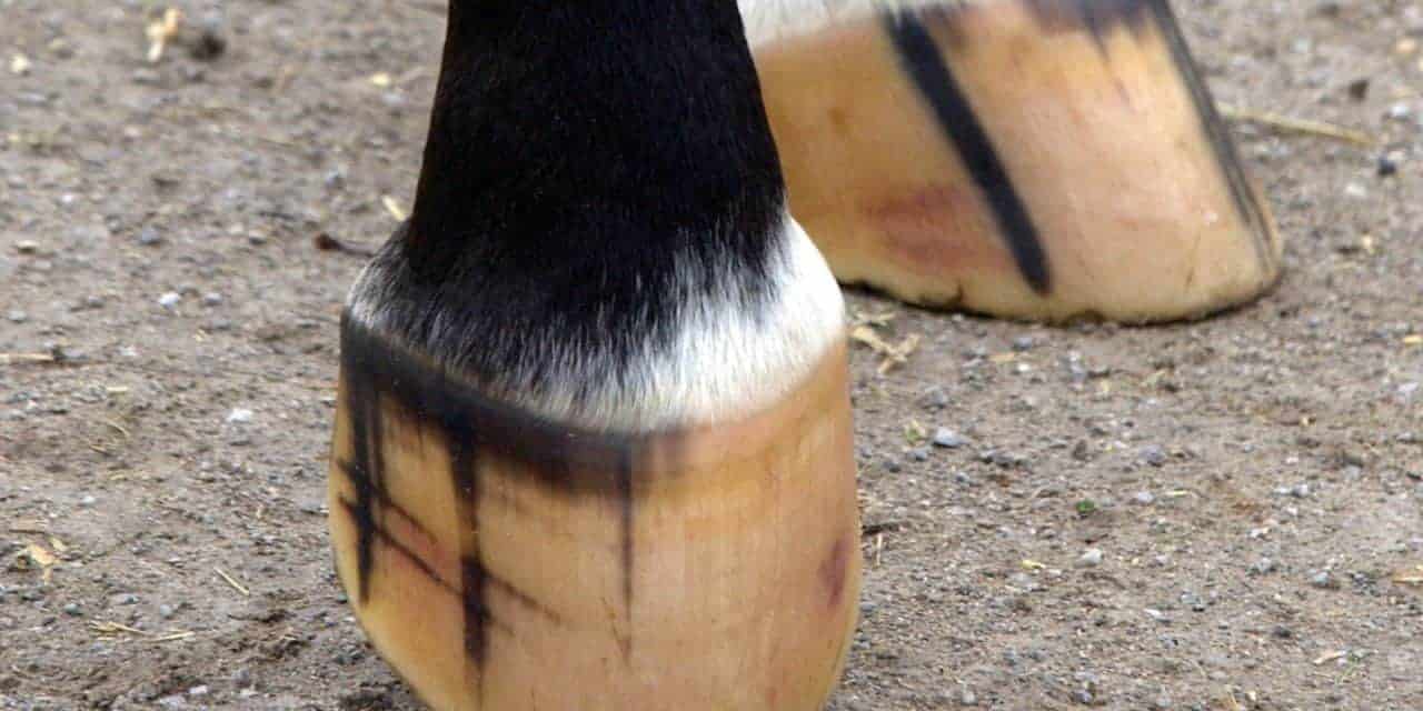
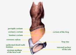
The coronary band is at the top of the hoof wall and is usually light in color. It is the growth and nutritional source for the hoof wall and contains a large supply of blood. If the coronary band is injured it can result in damage to the hoof wall and impeded hoof growth to the point where the horse may no longer be able to be ridden.
Peripople
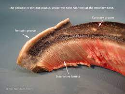
The periople covers the area just below the coronary band, protects the hoof wall. It is made up of soft newly formed hoof wall tissue, and helps give the hoof time to harden.
Inner Wall or Laminar Layer
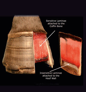
The inner wall of the hoof is more pliable than the hard outer wall. allowing the inner wall to expand with movement and act as a shock absorb-er, protecting the interior of the hoof. The laminae attaches the coffin bone to the inside of the hoof wall, and bears much of the horses’s weight.
Under the Hoof
Sole
The sole is the underside of the hoof, and helps to protect the inner workings of the hoof. Most of it does not make contact with the ground because it is a bit concave. The structure of the sole is similar to that of the hoof wall, and bears the internal weight that is transferred through the border of the sole rather than weight from the ground. An important area of the sole is the “white line.” The white line is located between the hoof wall and the sole. It’s tissues protect and help adhere the sole to the inner wall of the hoof.
Frog
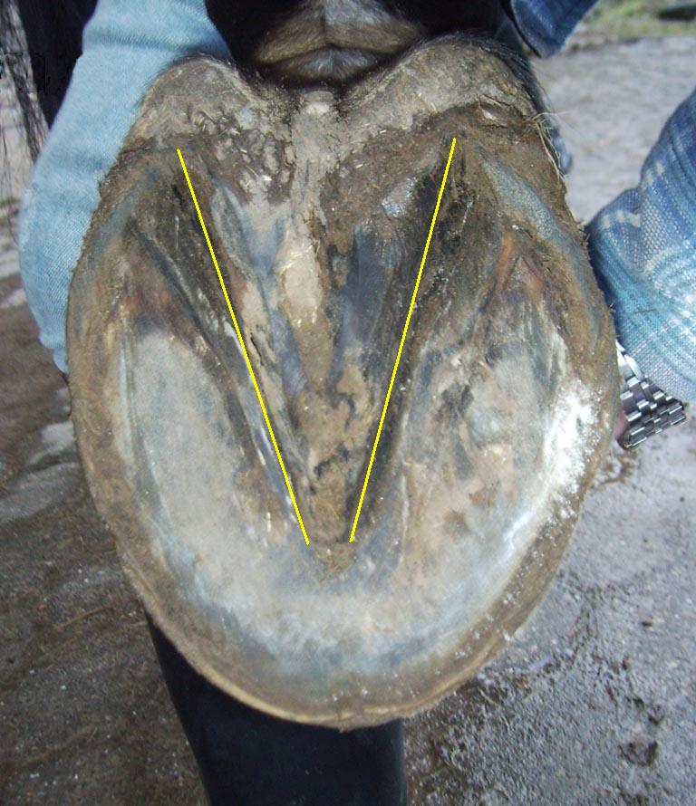
The frog is the tough, thick, V-shaped structure pointing to the front of the hoof. It’s function is to protect the cushion beneath it, and aid in traction and circulation in the hoof. It acts as shock absorber when the horse moves. The sensitive nerves helps the horse know where his feet are, and helps him feel the surface beneath him.
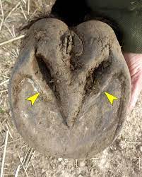
Central Sulcus
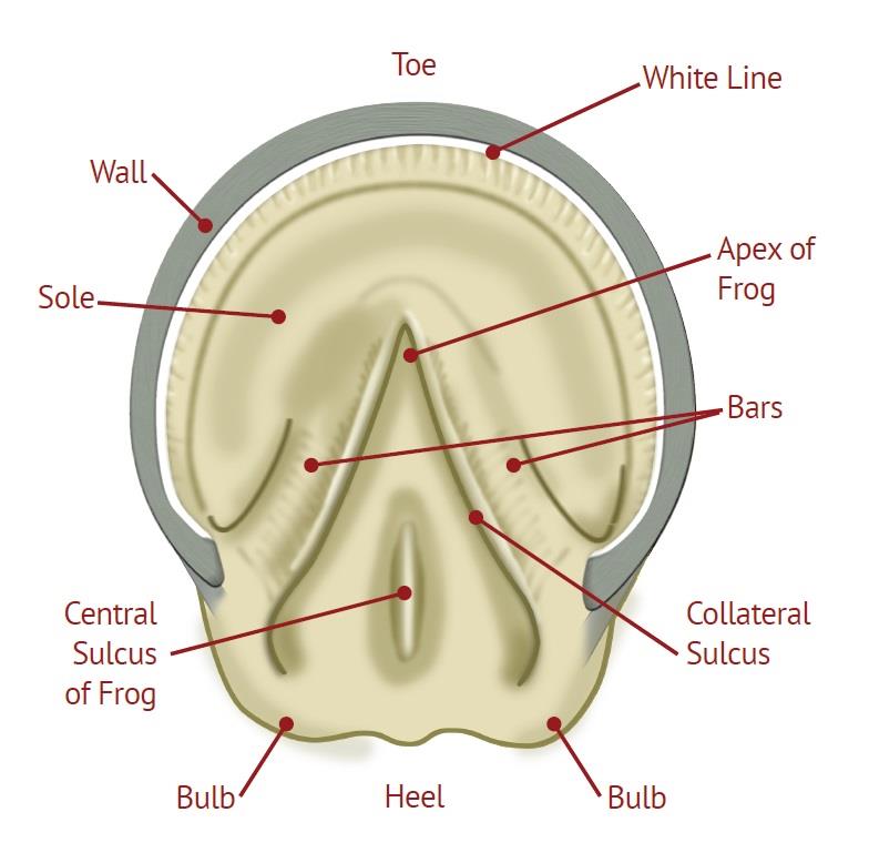 The groove down the center is the central sulcus, and the grooves on either side of the frog are the central and lateral sulci. The central sulcus should be fairly wide and shallow.
Bars
The bars are extensions of the hoof wall that strengthen the heel area and control over-expansion of the heels. They help with building the sole of the hoof and helps support the horse’s weight.
Interior Framework
Digital Cushion

The digital cushion is a cushion of cartilaginous material with some “give,” acting as one of the main shock absorbers in the hoof.
Coffin Bone
The coffin bone is located at the bottom near the toe and encapsulated in the hoof. It is the largest bone in the hoof and is surrounded by tissues that help make-up the laminae of the hoof wall, and the tissues of the sole.
Navicular Bone
The navicular bone is the small bone behind the coffin bone and the pastern bone. It helps stabilize the coffin bone and allows for some flexibility on uneven ground. Two tendons support and move the bones; the extensor tendon and the flexor tendon. The extensor tendon is attached to the coffin bone and straightens the leg; the flexor tendon runs down the back of the leg and wraps around the navicular bone, bending and flexing the leg.
Hopefully this gives you a good overview of hoof anatomy. Although with horses, you never know what’s going to happen… if you keep your horse’s hooves healthy with regular trims, a good diet and plenty of exercise you’re doing your part!
Subscribe to The Barngoddess Chronicles for more great articles on horses and equestrian life!